Evaluation of a Single Diagnostic Platform to Test Three Vibrio Species (Vibrio parahaemolyticus, Vibrio vulnificus and toxigenic Vibrio cholerae) in White Shrimp and Tiger Prawn for Field Monitoring and Laboratory Confirmation
Hwei Yih Pang*, Hilary Ee Lynn Boo and Angelito O. Abaoag
*Corresponding author: Hwei Yih Pang, Microbiome Life Sciences Pte Ltd, 150 South Bridge Road, #11-04 Fook Hai Building, Singapore. Email: pangkatrinna@mlsciences.com
DOI: 10.37722/JAMBE.2026302
Abstract
Vibrio species, including Vibrio parahaemolyticus, Vibrio vulnificus, and toxigenic Vibrio cholerae, are important bacterial pathogens associated with shrimp aquaculture and seafood safety. Routine monitoring is often limited by the lack of diagnostic platforms that can be applied consistently across field and laboratory settings. This study evaluated a unified diagnostic workflow based on a single TaqMan probe assay that can be used for both PCR screening and laboratory qPCR confirmation.
A total of 100 seafood samples comprising 50 white shrimp (Litopenaeus vannamei) and 50 tiger prawns (Penaeus monodon) were collected from five wet markets in Singapore. DNA was extracted using a rapid Chelex-based method and analyzed using a three-tube assay targeting the toxR, vvhA, and ctxA genes of V. parahaemolyticus, V. vulnificus, and toxigenic
V. cholerae, respectively. Amplification was performed using four instruments comprising two PCR systems and two qPCR systems. Diagnostic agreement between the PCR and qPCR systems was evaluated using Cohen’s kappa analysis.
Watch the Article in Motion
Authors:

Hwei Yih Pang
The assays demonstrated amplification efficiencies of 90.1–96.7% with limits of detection ranging from 14.25 to 15.67 copies per reaction. Overall agreement between the PCR and
qPCR systems ranged from 94.3% to 96.0%. Cohen’s kappa values ranged from 0.88 to 0.92, indicating almost perfect agreement according to the Landis and Koch (1977) interpretation scale. The highest agreement was observed between the Mini8/Mini16 (MiniPCR bio) and CFX96 (Bio-Rad) systems (κ = 0.92), whereas the lowest was observed between the Turbo Cycler (Blue-Ray Biotech) and Maverick (Anitoa) systems (κ = 0.88).
These findings demonstrate that a single assay design can provide reliable field-based qualitative screening and laboratory-based quantitative confirmation without requiring separate diagnostic workflows. The proposed diagnostic platform offers a practical approach for rapid Vibrio surveillance in shrimp and prawn production systems and supports timely disease monitoring and food safety management.
Keywords
Vibrio parahaemolyticus, Vibrio vulnificus, Vibrio cholerae, Litopenaeus vannamei, Penaeus monodon, Portable PCR, Quantitative PCR (qPCR), diagnostic concordance, Cohen’s kappa.
Introduction
Vibrio species such as Vibrio parahaemolyticus, Vibrio vulnificus, and Vibrio cholerae are of concern in shrimp and prawn farming. They are naturally present in aquatic environments and can rapidly proliferate under favorable conditions. This can lead to disease outbreaks, resulting in significant economic losses and posing risks to food safety and public health [1, 2, 3, 4]. To control disease outbreaks, routine surveillance is important for early intervention. Current monitoring approaches, including conventional microbiological culture, on-site screening methods, and testing by centralized laboratories, have several limitations.
Conventional microbiological culture methods remain widely used for the presumptive detection of Vibrio species. They are time-consuming, labor-intensive, and may underestimate bacterial presence when cells enter a viable but non-culturable (VBNC) state [5, 6, 7]. Field-deployable tests provide rapid results but may have limitations related to sample preparation, analytical sensitivity, and validation under field conditions [8]. While these tests offer the advantage of on-site detection, studies evaluating their agreement with laboratory-based qPCR remain limited. The common practice is to send samples to a centralized laboratory for qPCR testing, which is highly sensitive and reliable but requires sample transportation and longer turnaround times [9]. These logistical requirements may affect sample quality and delay decision-making, particularly when testing facilities are located far from the sampling site.
To address these limitations, we developed a unified diagnostic workflow based on a single TaqMan probe assay that can be used for both on-site screening and confirmatory quantification. During amplification, probe cleavage by DNA polymerase generates a fluorescent signal [10, 11]. This study used FAM-labelled probes because their fluorescence is compatible with the optical configuration of the P51 fluorescence viewer, enabling direct visualisation of amplified products without agarose gel electrophoresis while also supporting qPCR-based quantification [12]. This allows the same assay chemistry to be applied for both field-based qualitative screening and laboratory-based quantitative confirmation. Agreement between the two approaches was evaluated through a concordance study involving two PCR instruments and two qPCR instruments using Cohen’s kappa analysis [13, 14].
Materials and Methods
Sample collection and preparation
A total of 100 seafood samples, comprising 50 white shrimp (Litopenaeus vannamei) and 50 tiger prawns (Penaeus monodon), were collected from five wet markets across the North, Central, West, East, and North-East regions of Singapore between March and May 2026. Samples were purchased directly from retail vendors under normal consumer conditions to reflect typical market exposure. At each wet market, approximately 500 g of raw white shrimp and 500 g of raw tiger prawns were purchased based on product availability at the time of sampling. Species identity was determined based on vendor labeling. No additional ice or cooling was applied during transport. All samples were transported to the laboratory and processed on the same day of collection.
From each purchased batch, 10 individual shrimp or prawns were randomly selected for analysis, resulting in a total of 50 samples per species. Each shrimp or prawn was treated as an individual sample. The head was first removed using sterile scissors. The body tissue, including the gastrointestinal tract, was aseptically dissected, cut into small pieces, and transferred into sterile 50 mL Falcon tubes (Falcon, USA) containing 30 mL of phosphate-buffered saline (PBS; Ultra Pure Grade, 1st BASE). The tubes were gently inverted three to five times to mix the tissue with the buffer. The suspension was incubated at room temperature for 30–45 min to allow bacterial release into the buffer prior to DNA extraction for Vibrio detection.
DNA extraction
DNA was extracted using a rapid Chelex-based method. A 10% Chelex 100 Resin suspension (molecular biology grade, 200–400 mesh, sodium form; Bio-Rad, USA) was prepared. A 500 µL aliquot of the bacterial suspension was transferred to a
1.5 mL microcentrifuge tube (BIOFIL) and centrifuged at 8000 rpm for 10 min. The supernatant was discarded, and the pellet was resuspended in 100 µL of 10% Chelex solution by thorough pipetting. The mixture was transferred to a 0.2 mL PCR tube (Axygen) and heated at 95°C for 20 min using a Turbo Cycler TCST-9622 (Blue-Ray Biotech). After heating, the tubes were centrifuged at 2000 rpm for 1 min. The DNA-containing supernatant was carefully transferred to a clean 0.2 mL PCR tube while avoiding carryover of the Chelex resin. The extracted DNA was stored at −20°C until use. For all PCR and qPCR assays, 2 µL of extracted DNA was used as the template.
PCR and qPCR Assay
Information on the target genes for the three Vibrio species is summarized in Table 1. Primers were synthesized in Lab Ready format, and hydrolysis probes were synthesized as double-quenched probes labeled with ZEN/Iowa Black™ (Integrated DNA Technologies, IDT, USA). PCR and qPCR assays were performed in a 20 µL reaction volume using SensiFAST™ No-ROX Probe Master Mix (Bioline), with 2 µL of extracted DNA as the template. Thermal cycling conditions were standardized across all instruments, comprising an initial denaturation at 95°C for 5 min, followed by 45 cycles of denaturation at 95°C for 10 s and combined annealing/extension at 60°C for 20 s.
Amplification was performed using four instruments: two PCR systems, the miniPCR mini8 and mini16 Thermal Cyclers (MiniPCR bio, USA) and the Turbo Cycler TCST-9622 (Blue-Ray Biotech Corp., Taiwan), and two qPCR systems, the Maverick MQ8 Portable qPCR System (Anitoa Systems, USA) and the CFX96 Touch Real-Time PCR Detection System (Bio-Rad Laboratories, USA).
PCR amplicons were visualized using a P51 fluorescence viewer (miniPCR bio) under blue light excitation (450–480 nm) with an orange filter for visualisation. Synthetic DNA targets (toxR, vvhA, and ctxA; 4000 copies/µL; Integrated DNA Technologies, IDT, USA) served as positive controls, while Chelex extraction blanks were used as negative controls.
| Target | Target Gene | Amplicon Length | Genbank Accession | Amplification Efficiency (%) | LOD Copies/ reaction | Cq value at LOD |
| Vibrio parahaemolyticus | toxR | 147 bp | CP114186.1 | 90.1 | 14.69 | 35.95 |
| Vibrio vulnificus | vvhA | 179 bp | CP126699.1 | 92.2 | 14.25 | 37.79 |
| Toxigenic Vibrio cholerae | ctxA | 176 bp | CP189277.1 | 96.7 | 15.67 | 36.03 |
Table 1: Analytical performance of diagnostic assay for Vibrio spp. targeting toxR (V. parahaemolyticus), vvhA (V. vulnificus) and ctxA (toxigenic V. cholerae) (Pang HY, unpublished).
Detection and result interpretation
End-point PCR products generated using the Mini8/Mini16 (MiniPCR bio) and Turbo Cycler (Blue-Ray Biotech) were visualized using a P51 fluorescence viewer (Amplyus Ltd., USA). Samples exhibiting visible fluorescence were interpreted as positive. For qPCR, amplification data were analyzed using the instrument software of the Maverick (Anitoa) and CFX96 (Bio-Rad). A cycle quantification (Cq) cut-off value of 40 was applied. Samples with Cq ≤ 40 were considered positive, whereas samples with no amplification or Cq > 40 were considered negative.
Results
Detection of Vibrio targets
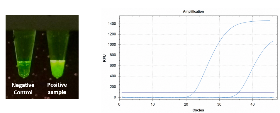
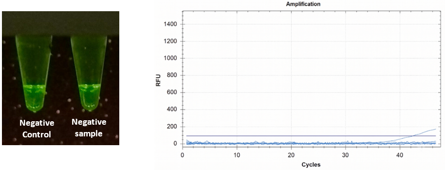
Representative positive results for Vibrio targets obtained by PCR and qPCR are shown in Figure 1. Representative negative samples are as shown in Figure 2.
Agreement between field PCR and laboratory qPCR systems
To evaluate agreement between PCR and qPCR systems, qualitative results from 300 reactions covering three Vibrio species were compared. Cohen’s kappa was calculated to measure agreement [14], and the Landis and Koch interpretation scale was used to classify the level of concordance [15].
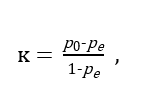
Cohen’s kappa formula:
The Landis & Koch Interpretation Scale 1977
< 0.00 : Poor agreement (less than chance)
0.01 – 0.20 : Slight agreement
0.21 – 0.40 : Fair agreement
0.41 – 0.60 : Moderate agreement
0.61 – 0.80 : Substantial agreement
0.81 – 1.00 : Almost perfect agreement
Table 2 shows the confusion matrix for four combinations of PCR and qPCR systems. Concordance analysis demonstrated
where,
almost perfect agreement across all evaluated platforms (Table 3). The highest level of concordance was observed between the Mini8/Mini16 (MiniPCR bio) vs CFX96 (Bio-Rad), yielding a Cohen’s kappa (κ) coefficient of 0.92 (95% CI: 0.879–0.961). The lowest, yet still robust, level of agreement occurred between the Turbo Cycler (Blue-Ray Biotech) vs Maverick (Anitoa) with κ = 0.88 (95% CI: 0.831–0.929). Overall, all
p0 : Observed Agreement. The actual percentage of times the raters agree. Calculated as the total number of agreements divided by the total number of ratings.
pe : Expected Chance Agreement. The hypothetical probability of chance agreement, calculated using the raters’ individual probabilities of assigning each category.
PCR/qPCR comparisons achieved κ values ≥ 0.88 with tight 95% confidence intervals, supporting the reliability and equivalence of both the PCR systems against standard qPCR frameworks.
| 1) | qPCR System: Maverick (Anitoa) | Total | ||
| – | + | |||
| PCR System: Mini8/Mini16 (MiniPCR bio) | – | 110 | 6 | 116 |
| + | 8 | 176 | 184 | |
| Total | 118 | 182 | 300 | |
| 2) | qPCR System: CFX96 (Bio-Rad) | Total | ||
| – | + | |||
| PCR System: Mini8/Mini16 (MiniPCR bio) | – | 111 | 5 | 116 |
| + | 7 | 177 | 184 | |
| Total | 118 | 182 | 300 | |
| 3) | qPCR System: Maverick (Anitoa) | Total | ||
| – | + | |||
| PCR System: Turbo Cycler (Blue-Ray Biotech) | – | 109 | 8 | 117 |
| + | 9 | 174 | 183 | |
| Total | 118 | 182 | 300 | |
| 4) | qPCR System: CFX96 (Bio-Rad) | Total | ||
| – | + | |||
| PCR System: Turbo Cycler (Blue-Ray Biotech) | – | 110 | 7 | 117 |
| + | 8 | 175 | 183 | |
| Total | 118 | 182 | 300 | |
Table 2: Confusion Matrix:
Mini8/Mini16 (MiniPCR bio) vs Maverick (Anitoa)
Mini8/Mini16 (MiniPCR bio) vs CFX96 (Bio-Rad)
Turbo Cycler (Blue-Ray Biotech) vs Maverick (Anitoa)
Turbo Cycler (Blue-Ray Biotech) vs CFX96 (Bio-Rad)
| PCR/qPCR Comparison | Agreement (%) | Cohen’s kappa, ᴋ | Standard Error, SE | 95% Confidence Interval, CI | Interpretation (Landis and Koch 1977) |
| 1 | 95.3 | 0.90 | 0.026 | 0.852 to 0.952 | Almost perfect agreement |
| 2 | 96.0 | 0.92 | 0.024 | 0.869 to 0.963 | |
| 3 | 94.3 | 0.88 | 0.028 | 0.826 to 0.936 | |
| 4 | 95.0 | 0.90 | 0.026 | 0.843 to 0.947 |
Table 3: Agreement analysis between PCR and qPCR system.
Mini8/Mini16 (MiniPCR bio) vs Maverick (Anitoa)
Mini8/Mini16 (MiniPCR bio) vs CFX96 (Bio-Rad)
Turbo Cycler (Blue-Ray Biotech) vs Maverick (Anitoa)
Turbo Cycler (Blue-Ray Biotech) vs CFX96 (Bio-Rad)
Disagreement between field PCR and laboratory qPCR systems
A total of 27 discordant sample-target combinations were identified across all PCR and qPCR platform comparisons. Of these, 25 were associated with V. vulnificus and 2 with V. parahaemolyticus, while no discordant results were observed for toxigenic V. cholerae.
Discussion
Degree of PCR and qPCR Agreement
Cohen’s kappa incorporates the possibility of agreement occurring by chance and is commonly used for diagnostic concordance studies involving categorical positive or negative outcomes. The Cohen’s kappa results indicate strong qualitative agreement beyond chance. Based on the interpretation criteria proposed by Landis and Koch, all platform comparisons were classified as demonstrating almost perfect agreement [15]. These findings indicate high qualitative consistency between portable PCR systems used for field screening and laboratory-based qPCR systems used for confirmatory analysis.
Most discordant observations involving V. vulnificus were associated with low level target detection, with corresponding qPCR Cq values ranging from Cq 35.13 to Cq 39.98, with the mean Cq 38.1, which is close to the assay limit of detection (LOD Cq = 37.79). These findings suggest that a substantial proportion of the observed discrepancies were attributed to low target concentration and stochastic effects near the detection threshold, rather than to systematic differences between PCR and qPCR systems [16]. Only two discordant observations were associated with V. parahaemolyticus (samples B07S and E10P). Notably, sample B07S yielded relatively low qPCR Cq values (20.03–20.94), indicating that factors other than target concentration may have contributed to this isolated discrepancy.
Despite these occasional discordant results, all platform comparisons demonstrated almost perfect agreement based on Cohen’s kappa analysis (Table 3), supporting the suitability of the proposed workflow for both field screening and laboratory confirmation.
The use of Chelex Extraction Method
Chelex-based extraction provides a rapid and reliable method for nucleic acid preparation in aquatic pathogen detection. Compared with conventional commercial extraction kits, the Chelex extraction method requires fewer processing steps, shorter extraction time, lower cost, less technical expertise and less equipment, making it more suitable for portable or resource-limited testing conditions [17, 18]. Although Chelex extraction may produce DNA of lower purity and retain higher levels of potential PCR inhibitors, previous studies have demonstrated that it provides acceptable amplification performance [17, 19].
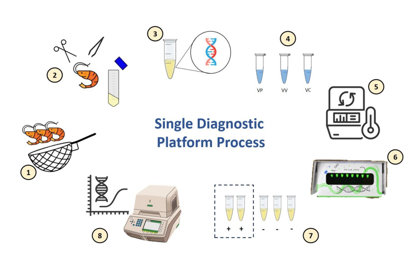
Proposed Single Diagnostic Platform Process
1) Sampling (approx. 30 min): Fresh shrimp and prawn samples were collected and processed immediately on-site to preserve DNA integrity and minimize transport delays.
2) Sample processing (approx. 1 hour): Tissues were dissected and homogenized in PBS buffer to prepare sample suspensions for DNA extraction.
3) DNA extraction (approx. 1 hour): DNA was extracted from the prepared suspensions using a rapid Chelex-based method.
4) Assay preparation (approx. 30 min): Extracted DNA was directly added to PCR reaction mixtures targeting Vibrio parahaemolyticus, Vibrio vulnificus, and toxigenic Vibrio cholerae.
5) PCR amplification (approx. 1.5 hour): Amplification was performed using portable thermocycler systems under standardized conditions.
6) Fluorescence detection (approx. 5 min): Amplification products were visualized using a portable blue-light viewer, where fluorescence indicated target presence.
7) Transport of positive samples (distance-dependent): Only positive DNA extracts were transported to the laboratory for confirmatory analysis, reducing transport load and contamination risk.
8) qPCR confirmation (approx. 1.5 hour): Positive extracts were directly subjected to qPCR for confirmation and quantification based on Cq values.
Advantages and Limitations in Vibrio Surveillance
Routine surveillance is a fundamental component of disease prevention in shrimp and prawn aquaculture because pathogenic Vibrio species can persist in the aquatic environment and colonize animals before clinical signs become evident [20, 21]. Beyond supporting early outbreak detection and management, surveillance provides essential data for risk assessment and policy development while reducing the likelihood of underestimating the presence and distribution of pathogenic Vibrio [20]. The proposed diagnostic platform provides a practical tool for routine farm-level monitoring and disease prevention throughout the aquaculture production chain, from broodstock and hatchery screening to nursery, grow-out ponds, harvest, and post-harvest seafood testing. Its portability enables more frequent on-site testing than conventional laboratory-based approaches, facilitating timely pathogen detection, early biosecurity interventions, and preventive disease management.
Multiplex PCR and qPCR assays enable the simultaneous detection of multiple Vibrio species in a single reaction, reducing assay time and overall reagent consumption. However, the inclusion of multiple primer sets in a single reaction may introduce challenges such as target competition, altered amplification efficiency, primer–dimer formation and non-specific amplification, often requiring extensive assay optimization [22, 23, 24]. In contrast, the present workflow employs separate singleplex assays under a common thermal cycling protocol, avoiding these potential interactions while maintaining a simple and unified diagnostic workflow. Furthermore, because each target is amplified in an individual reaction, fluorescence signals can be interpreted independently during endpoint visual inspection, simplifying result interpretation.
Portable molecular methods such as loop-mediated isothermal amplification (LAMP) and recombinase polymerase amplification (RPA) have been widely explored for rapid field detection of Vibrio species. However, studies directly comparing these methods with qPCR remain limited. Available studies have reported higher analytical sensitivity for qPCR than LAMP or real-time LAMP despite comparable diagnostic performance [25-27]. In contrast, real-time RPA demonstrated complete concordance with qPCR for Vibrio detection, although this agreement was established using positive samples with qPCR Ct values ≤ 26.13, and its performance at higher Ct values remains unclear [28]. Furthermore, because LAMP and RPA employ amplification mechanisms distinct from PCR and qPCR, they require independent assay development and validation. In contrast, the proposed workflow uses a single TaqMan probe assay compatible with both PCR and qPCR, enabling seamless transition from field screening to laboratory confirmation using the same molecular assay. A limitation of the endpoint fluorescence PCR assay is that it provides only qualitative results and cannot estimate target abundance. Therefore, it is intended as a rapid field screening tool, with qPCR using the same assay providing quantitative confirmation when required.
The endpoint fluorescence PCR assay demonstrated high qualitative agreement with the corresponding qPCR assay, supporting its application as a reliable field-screening tool. Although its analytical limit of detection was not formally evaluated, the present findings support diagnostic agreement rather than equivalent analytical sensitivity. Future studies should determine the analytical limit of detection through serial dilution experiments and directly compare its analytical performance with other portable amplification methods under identical experimental conditions.
Improvement and Recommendation
The initial field-based screening approach was designed to provide rapid qualitative detection of target Vibrio species but a positive result does not necessarily indicate an active disease outbreak. It is because they are natural microflora in shrimp and prawn aquaculture environments, and their detection may occur even in clinically healthy samples [29]. Consequently, diagnostic results should be interpreted within a broader epidemiological context, including bacterial load, animal health status, environmental parameters, and farm conditions, to more accurately assess disease risk. Based on these combined field and farm-level assessments, a Cq threshold for disease risk classification can be established and refined.
Establishing a Cq threshold for disease risk would enable the assay to be advanced beyond a simple presence–absence test into a practical and quantitative risk screening tool. This approach aligns with quantitative microbial risk assessment principles, where molecular detection outputs are translated into meaningful risk categories for decision making [30]. Such refinement may be supported through optimisation of assay parameters to improve control of fluorescence signal intensity in PCR detection. The agreement between PCR-based results and the proposed Cq-based risk classification could be evaluated using Cohen’s kappa analysis.
The present study was conducted using samples collected from a limited number of wet markets in Singapore and may not fully represent the broader diversity of Vibrio contamination patterns across different aquaculture systems or geographic regions. A larger sample size would be required to improve the robustness of the findings [31].
Conclusion
Overall, the proposed workflow demonstrates a practical approach for rapid detection of multiple Vibrio species by combining endpoint fluorescence PCR for field screening with laboratory confirmatory qPCR across the aquaculture production chain. Although each target is detected using a separate species-specific assay, the shared thermal cycling conditions allow multiple Vibrio species to be tested within the same PCR run without requiring separate amplification protocols. This simplifies routine surveillance, enabling earlier detection of pathogenic Vibrio species, timely diagnostic decision-making, and more effective biosecurity interventions. In addition, rapid screening of both aquaculture samples and seafood products supports food safety by enabling the early detection of foodborne Vibrio pathogens before contaminated products enter the consumer market. With further validation, this workflow may also provide a foundation for the development of risk-based surveillance strategies to support aquaculture health management.
Declaration of competing interest
The authors declare that they have no known competing financial interests or personal relationship that could have appeared to influence the work reported in this paper.
References
- Nguyen HT, Van TN, Ngoc TT, Boonyawiwat V, Rukkwamsuk T, Yawongsa A (2021). Risk factors associated with acute hepatopancreatic necrosis disease at shrimp farm level in Bac Lieu Province, Vietnam. Vet World. 14(4):1050-1058. https://pmc.ncbi.nlm.nih.gov/articles/PMC8167526/
- Kumar V, Roy S, Behera BK, Bossier P, Das BK (2021). Acute Hepatopancreatic Necrosis Disease (AHPND): Virulence, Pathogenesis and Mitigation Strategies in Shrimp Aquaculture. Toxins (Basel) 13(8):524. https://pubmed.ncbi.nlm.nih.gov/34437395/
- EFSA BIOHAZ Panel (EFSA Panel on Biological Hazards), Koutsoumanis K, Allende A, Alvarez-Ordóñez A, Bolton D, et al (2024). Public health aspects of Vibrio spp. related to the consumption of seafood in the EU. EFSA Journal 22: 7. https://efsa.onlinelibrary.wiley.com/doi/10.2903/j.efs a.2024.8896
- Jara-Medina NR, Cedeño-Pinargote AC, Beltrán-Noboa A, Tejera E, Machado A (2025). Managing Vibrio parahaemolyticus and Vibrio alginolyticus Infections in the Whiteleg Shrimp (Penaeus vannamei): A Systematic Review. Molecules 30(17):3620. https://pubmed.ncbi.nlm.nih.gov/40942145/
- Oliver JD (2010). Recent findings on the viable but nonculturable state in pathogenic bacteria. FEMS Microbiology Reviews 34(4): 415-25. https://doi.org/10.1111/j.1574-6976.2009.00200.x
- Fakruddin M, Mannan KS, Andrews S (2013). Viable but nonculturable bacteria: Food safety and public health perspective. ISRN Microbiol. 2013: 703813. https://pmc.ncbi.nlm.nih.gov/articles/PMC38 04398/
- Li L, Mendis N, Trigui H, Oliver JD and Faucher SP (2014). The importance of the viable but non-culturable state in human bacterial pathogens. Front. Microbiol. 5:258. https://www.frontiersin.org/journals/microbiology/art icles/10.3389/fmicb.2014.00258/full
- Liu Q, Qiu Z, Yao M, Jiao B, Zhou Y, Li C, Liu H, Xin L (2026). Progress of Rapid Detection Technology for Aquatic Microorganisms: A Comprehensive Review. Microorganisms. 14(4):939. https://www.mdpi.com/2076-2607/14/4/939
- Zhang B, Qiu Y, Shi C, Zhang J (2025). Development of Multiple Real-Time Fluorescent Quantitative PCR for Vibrio Pathogen Detection in Aquaculture. Veterinary Sciences 12(4):327. https://www.mdpi.com/2306-7381/12/4/327
- Holland PM, Abramson RD, Watson R, Gelfand DH (1991). Detection of specific polymerase chain reaction product by utilizing the 5′ 3′ exonuclease activity of Thermus aquaticus DNA polymerase. Proc Natl Acad Sci USA. 88(16):7276-80. https://pmc.ncbi.nlm.nih.gov/articles/PMC52277/
- Heid CA, Stevens J, Livak KJ, and Williams PM (1996). Real time quantitative PCR. Genome Research 6: 986-994. https://genome.cshlp.org/content/6/10/986
- Misquitta K, Miller BM, Malecek K, Gleason E, et al (2024). A fluorescence viewer for rapid molecular assay readout in space and low-resource terrestrial environments. PLoS One 19(3):e0291158. https://pmc.ncbi.nlm.nih.gov/articles/PMC10942025/
- Cohen J (1960). A coefficient of agreement for nominal scales. Educational and Psychological Measurement 20(1):37–46. https://journals.sagepub.com/doi/10.1177/001316446 002000104
- McHugh ML (2012). Interrater reliability: the kappa statistic. Biochem Med (Zagreb) 22(3):276-82. https://pubmed.ncbi.nlm.nih.gov/23092060/
- Landis JR, Koch GG (1977). The measurement of observer agreement for categorical data. Biometrics 33(1):159-74. https://pubmed.ncbi.nlm.nih.gov/843571/
- Bustin SA, Kirvell S, Nolan T, Mueller R, Shipley GL (2025). When Two-Fold Is Not Enough: Quantifying Uncertainty in Low-Copy qPCR. Int J Mol Sci. 26(16):7796. https://pmc.ncbi.nlm.nih.gov/articles/PMC12387016/
- Hu X, Jiang N, Li Y, Zhou Y, Fan Y, Xue M, Zeng L, Liu W, Meng Y (2022). Rapid Nucleic Acid Extraction for Aquatic Animal DNA Virus Determination Using Chelex 100 Resin via Conventional PCR and Digital Droplet PCR Detection. Animals (Basel). 12(15):1999. https://pubmed.ncbi.nlm.nih.gov/35953988/
- Walsh PS, Metzger DA, Higuchi R (1991). Chelex 100 as a medium for simple extraction of DNA for PCR-based typing from forensic material. Biotechniques 10(4):506-13. https://pubmed.ncbi.nlm.nih.gov/1867860/
- Yang H, Zhou Q, Hu J, Bao Z, Wang M (2024). A Chelex-100-based rapid DNA extraction method and its application in the detection of shrimp pathogens. Electronic Journal of Biotechnology 70:29-37. https://www.sciencedirect.com/science/article/pii/S07 17345824000125
- Hobe A, Msolo L, Ebomah KE, Jaja IF, Oguttu JW, Okoh AI (2026). Antibiotic Resistance and Pathogenicity of Vibrio Species in Aquaculture: Implications for Fish Health and Food Safety in LMICs. Vet Med Sci. 12(2):e70877. https://pubmed.ncbi.nlm.nih.gov/41830177/
- Vandeputte M, Kashem MA, Bossier P, Vanrompay D (2024). Vibrio pathogens and their toxins in aquaculture: A comprehensive review. Rev Aquac. 16(4):1858-1878. https://onlinelibrary.wiley.com/doi/abs/10.1111/raq.1 2926
- Loo KY, Law JWF, Tan LTH, Pusparajah P, Letchumanan V, Lee LH (2022). Diagnostic techniques for rapid detection of Vibrio species. Aquaculture 561: 738628.https://www.sciencedirect.com/science/article/pii/S00 4484862200744X
- Toohey-Kurth K, Reising MM, Tallmadge RL, Goodman LB, Bai J, Bolin SR, Pedersen JC, Bounpheng MA, Pogranichniy RM, Christopher-Hennings J, Killian ML, Mulrooney DM, Maes R, Singh S, Crossley BM (2020). Suggested guidelines for validation of real-time PCR assays in veterinary diagnostic laboratories. J Vet Diagn Invest. 32(6):802-814. https://pubmed.ncbi.nlm.nih.gov/32988335/
- Sint D, Raso L, Traugott M (2012). Advances in multiplex PCR: balancing primer efficiencies and improving detection success. Methods Ecol Evol. 3(5):898-905.https://pmc.ncbi.nlm.nih.gov/articles/PMC3573865/
- Telli AE, Doğruer Y (2019). Discrimination of viable and dead Vibrio parahaemolyticus subjected to low temperatures using Propidium Monoazide – Quantitative loop mediated isothermal amplification (PMA-qLAMP) and PMA-qPCR. Microbial Pathogenesis 132: 109-116. https://www.sciencedirect.com/science/article/abs/pii/S0882401018316024
- Cao X, Zhao LC, Zhang JF, Chen X, Shi L, Fang X, Xie H, Chang YL, Wang Li (2019) Detection of viable but nonculturable Vibrio parahaemolyticus in shrimp samples using improved real-time PCR and real-time LAMP methods. Food Control 103: 145-152.https://www.sciencedirect.com/science/article/abs/pii/S0956713519301422
- Jones JL, Hara-Kudo Y, Krantz JA, Benner RA Jr, Smith AB, Dambaugh TR, Bowers JC, Depaola A (2011). Comparison of molecular detection methods for Vibrio parahaemolyticus and Vibrio vulnificus. Food Microbiol. 30(1):105-11. https://pubmed.ncbi.nlm.nih.gov/22265290/
- Wang JX, Tang WL, Chen SQ, Zhang J, Ji J, Dong JQ, Liu G, Gao S (2021). Rapid and sensitive detection of Vibrio alginolyticus pathogenic strains by real-time recombinase polymerase amplification. Acta Biochimica et Biophysica Sinica 53(7):950-954. https://academic.oup.com/abbs/article/53/7/950/6237953
- Thompson FL, Lida T, Swings J (2004). Biodiversity of Vibrios. Microbiol Mol Biol Rev 68:3. https://journals.asm.org/doi/10.1128/mmbr.68.3.403-431.2004 Hartfield M, Alizon S (2017) Introducing the outbreak threshold in epidemiology. PLoS Pathog. 13(5):e1006415.https://pmc.ncbi.nlm.nih.gov/articles/PMC3680036/
- Hoelzer K, Pouillot R (2013). Practical Considerations for the Interpretation of Microbial Testing Results Based on Small Numbers of Samples. Foodborne Pathogens and Disease. 10(11):907-915. https://journals.sagepub.com/doi/abs/10.1089/fpd.201 3.1531